


AGIBOT Dynamics
Expanding into a New Procedure: AGIBOT System's First Bariatric Surgery Completed at Qilu Hospital
Qilu Hospital of Shandong University has recently completed a sleeve gastrectomy using the AGIBOT Endoscopic Surgical System. The procedure was led by Professor Sanyuan Hu, academic leader of the Department of General Surgery at the hospital, with the participation of Professor Shaozhuang Liu, Director of the Department of Bariatric and Metabolic Surgery. This marks the first bariatric surgery performed with the AGIBOT System. Robot docking took 5 minutes, the surgeon's console time was 74 minutes, and intraoperative blood loss was approximately 10 mL. The procedure went smoothly.
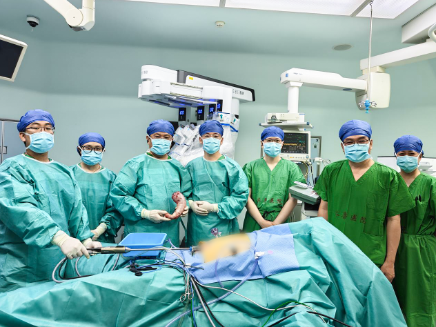
The AGIBOT System's first bariatric surgery completed successfully
Beyond Weight Loss: A Focus on Metabolic Improvement
The patient was a young male with a BMI above 35 kg/m². Previous attempts to lose weight through diet management and exercise had achieved unsatisfactory results, and he also presented with abnormalities in glucose metabolism, blood pressure and lipid metabolism. The surgery was performed to address the treatment needs of obesity combined with metabolic abnormalities. For bariatric and metabolic surgery, the value of the procedure is also reflected in multiple aspects, including glucose and lipid metabolism and liver health.
Metabolic dysfunction-associated steatohepatitis (MASH) is one area that has attracted attention. A randomized controlled trial has shown that sleeve gastrectomy can enable some patients with obesity and concurrent steatohepatitis to achieve histological resolution of steatohepatitis without worsening of fibrosis at one year after surgery [1]. The findings point to the therapeutic potential of bariatric and metabolic surgery for obesity-related liver disease, while individual benefits still need to be assessed in light of each patient's specific condition and long-term follow-up.
During surgery, treatment goals must be translated into precise control of the resection extent, tissue preservation and the morphology of the gastric sleeve. Sleeve gastrectomy resects most of the gastric body and fundus along the greater curvature of the stomach, preserves the tubular structure along the lesser curvature and the pylorus, and reshapes the originally broad stomach into a narrow "sleeve."
Gastric capacity needs to be reduced appropriately, while the remaining "sleeve" must maintain good blood supply, a straight configuration and an appropriate caliber. This requires the surgeon to fully expose the resection boundary during mobilization and to balance local width with overall alignment during fashioning, so that tissue preservation and sleeve construction are integrated throughout the continuous procedure.
Patient-Tailored Setup: Coordinated Three-Arm Operation
To operate from the greater curvature to the left subphrenic space, the instrument access paths must first be carefully planned. Taking the patient's body habitus into account, Professor Sanyuan Hu chose a three-arm configuration with subumbilical port placement, establishing operative paths in the upper abdomen. This setup enabled the instruments to reach the deep fundic region while accommodating the retraction and manipulation required for subsequent sleeve fashioning, and it left room for the patient-side assistant.
Once the setup was finalized, the team completed docking with the AGIBOT System in 5 minutes, after which the surgeon's console operation and patient-side coordination proceeded in tandem.
Meticulous Mobilization, Clearly Defined Resection Boundaries
Intraoperative blood loss in this case was approximately 10 mL. The identification and management of perigastric vessels is a key element of intraoperative bleeding control and places specific demands on the precision of exposure and dissection.
As the operation advanced toward the left subphrenic space, the gastric fundus became the region requiring meticulous handling. Located beneath the left hemidiaphragm and adjacent to the spleen, the fundus is connected to the spleen by the short gastric vessels, and its posterior attachments can restrict tissue deployment. To obtain a clear view of the fundus, the tissues must be fully deployed; yet the direction and extent of retraction directly bear on the protection of the adjacent spleen and vessels. Exposure and protection therefore need to be addressed within the same maneuver.
The 4K/3D vision of the AGIBOT System helped the surgeon identify tissue planes, vascular courses and instrument positions; its 7-degrees-of-freedom instruments could adapt to the subphrenic operating angles, adjust end-effector orientation and assist retraction and tissue deployment, allowing dissection and vascular management in the deep region to proceed under clear visualization.
Mobilization of the fundus also establishes the boundary for subsequent resection. Fully deploying the fundus and its posterior attachments helps clearly expose the gastroesophageal junction and determine the proximal resection extent. During dissection, the surgeon flexibly adjusted the direction and extent of instrument retraction through the AGIBOT System. The AGIBOT energy devices delivered appropriate output throughout this surgery, supporting tissue dissection and vascular management while balancing hemostasis with the protection of adjacent tissues.
Meticulous Sleeve Fashioning, Balancing Capacity and Patency
As the procedure entered the sleeve-fashioning phase, the surgeon's focus shifted from "how to deploy the tissues fully" to "how to preserve the proper configuration." Each transection had to be judged within the context of the final alignment of the gastric sleeve.
The angular incisure at the junction of the gastric body and antrum was a key transition point requiring particular attention. Excessively narrow local resection could compromise patency, while uneven retraction of the anterior and posterior gastric walls could cause twisting of the sleeve. Accordingly, the transection direction and retraction method at hand also influenced how the next segment of tissue would connect.
Under the three-dimensional vision of the AGIBOT System, the surgeon could adjust instrument retraction according to the deployment of the gastric wall, keep the anterior and posterior walls smooth and evenly tensioned, and judge the width to preserve and the direction of transection. Excessive narrowing was avoided when passing the angular incisure, and protection of the junctional tissues was maintained while advancing proximally, allowing each transected segment to connect naturally. After resection, the patency of the gastric sleeve was rechecked based on staple-line hemostasis, tissue blood supply and overall morphology.
For this surgery, reducing gastric capacity and preserving a healthy gastric sleeve were objectives that had to be achieved together; the ultra-HD vision and instrument flexibility of the AGIBOT System were integrated into the key steps of exposure, dissection and fashioning in service of these goals.
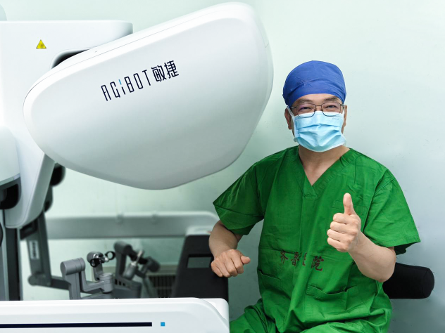
Professor Sanyuan Hu operating the AGIBOT System
After the surgery, Professor Sanyuan Hu commented: "Sleeve gastrectomy requires not only control of the resection extent, but also attention to the morphology and patency of the gastric sleeve. Whether the fundus can be fully exposed and whether balanced retraction can be maintained both affect the subsequent steps. In this case, the AGIBOT System provided clear vision and flexible adjustment of instrument angles, and its three-arm configuration met the operative demands of this surgery, with smooth overall coordination."
Previously, the team of Professor Sanyuan Hu had already used the AGIBOT System to perform procedures including spleen-preserving distal pancreatectomy and partial hepatectomy. The completion of this sleeve gastrectomy adds a new surgical procedure to the ongoing clinical applications of AGIBOT at Qilu Hospital of Shandong University.
Reference:
[1] Verrastro O, Panunzi S, Castagneto-Gissey L, et al. Bariatric-metabolic surgery versus lifestyle intervention plus best medical care in non-alcoholic steatohepatitis (BRAVES): a multicentre, open-label, randomised trial[J]. Lancet, 2023, 401(10390): 1786–1797. DOI: 10.1016/S0140-6736(23)00634-7.
View the original study (PubMed): https://pubmed.ncbi.nlm.nih.gov/37088093/
2026.09.23